How To Prevent Toenail Fungus
No one wants to experience toenail fungus (also called onychomycosis). However, it is relatively...
Strengthen your nails with Hydrate
Nail anatomy is very complex and the sensitivity of our fingertips are correlated to the complexity of the parts of the nail; however, only the following main parts of the nail will be discussed: the matrix, nail plate, nail bed, nail root, lunula, eponychium, and the paronychium.
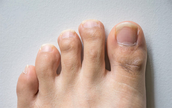
Starting at the top of the nail, the “nail plate” is the term for the actual nail, which is formed from layers of keratin protein from bonded amino acids. Due to its translucent nature, blood vessel pigments are visible, and the shape of the nail plate differs based on the underlying bone. Underneath the nail plate lies the “nail bed”, which is the skin tissue that contains blood capillaries and glands. The “matrix”, on the other hand, is the part of the nail bed that extends from the “nail root” and contains lymph cells, blood vessels, and nerves that play a role in the production of new cells for the nail. The “lunula” is the visible portion of the matrix and appears at the base of some fingernails with a white crescent shape. The “eponychium” is the skin tissue covering the new nail plate, also known as the “cuticle”, and the “paronychium” is the skin surrounding the sides of the nail, where hangnails may appear. Usually humans only possess one nail on each finger or toe tip; however, some people have inherited “sixth toenails” with a fifth toenail that is separated into two parts (2).
Healthy nails are usually pinkish to skin-colored, but can vary in shape and appearance according to the individual’s genetic makeup. The presence of melanocytes in the nail is relative to the amount of melanin within the skin, meaning more pigmented nails accompany darker skin. Similar to lighter skin, melanocytes only appear after serious injuries to the nail (e.g., slamming your finger in the car door) (1). Millions of blood vessels are present underneath the nail bed, which is why your nail turns dark purple due to the ruptured blood vessels (2).

Nail growth is a slow and gradual process, in which longer appendages grow faster than shorter ones. Fingernails grow three times faster than toenails, and growth among the fingernails is relative to length, with the middle finger growing the fastest at 3-4mm per month. In contrast, toenails grow slower at a rate of 1mm per month and may take up to 18 months to fully develop a new nail (1). Nail growth rate is determined by intrinsic factors (e.g., genetic and hereditary aspects, age, and gender) and also by extrinsic conditions (diet, exercise, and season) (2). In fact, nails tend to grow slower in the winter than in the summer due to the abundance of vitamin D from the sun. Since nail growth is stimulated by blood circulation throughout the capillaries, the nails halt their growth after you die. Nails may only appear longer after death because loss of water in the skin causes your cuticles and the surrounding skin to shrink back (2).
From an evolutionary point of view, nails can be considered as human “claws” that allow enhanced dexterity of the hands, such as scratching, clawing, picking, and pinching. Nails primarily serve as a source of protection for the tips of your fingers, which are medically termed “distal phalanges” (2). This area needs protection because thousands of nerve endings are concentrated within your fingertips, which serve as one of our main five senses – “touch”. Fingertips are used to determine whether our surroundings are safe, such as the temperature of surfaces, our food, and to serve as our “second eyes” in dark places or in those with compromised sight. The presence of the nail also serves to enhance the functional sensitivity of our fingernails by serving as straight edges for precision measurement (2). For example, telling the difference between a quarter and a dime would be difficult in the dark without your fingernails. In this case, you can think of your nails as tiny rulers! The nail also increases touch sensitivity due to its location over millions of open nerve endings and its ability to provide counter-pressure when you touch an object with the tip of your nail (2). This counterpressure stimulates the nerve endings underneath the nail bed.
1. Haneke, E. (2015). Anatomy of the nail unit and the nail biopsy. Seminars in Cutaneous Medicine and Surgery 34(2), pp. 95-100, doi: 10.12788/j.sder.2015.0143.
Retrieved from: https://www.researchgate.net/publication/280122178_Anatomy_of_the_nail_unit_and_the_nail_biopsy.
2. Semantic Scholar. Nail anatomy [PDF file]. Retrieved from: https://pdfs.semanticscholar.org/d55e/409902b393eefd4e20756cafdb9e8a560c08.pdf.